Skin Life & Aesthetic Clinic
Levels of alpha 1 anti-trypsin levels and respiratory functions in patients with pemphigus vulgaris
Levels of alpha 1 anti-trypsin levels and respiratory functions in patients with pemphigus vulgaris
Couldn't load pickup availability
|
Attention for spellings, sentences Dr Tahir Kamal designation Tables/ figures not mentioned in txt. Figures legend text to write below figures, Tables numbering |
Original Article
Levels of alpha 1 anti-trypsin levels and respiratory functions in patients with pemphigus vulgaris
|
|
Tahir Kamal, Shaiqa Mufti
Department of Dermatology, Post Graduate Medical Institute, Ameer-ud-din Medical College, Lahore General Hospital, Lahore.
|
|
Abstract |
Objective To examine the role of alpha 1 anti-Trypsin and FEV1 in patients with pemphigus vulgaris.
Methods A case-control research was carried out on 10 biopsy-proven PV patients and ten age-matched healthy controls. Both groups had pulmonary function tests (FEV1, total lung capacity, vital capacity), as well as alpha 1-antitrypsin levels. The Pemphigus Disease Area Index (PDAI) was utilized to assess disease severity in the PV group. The results were examined using the One-Sample Kolmogorov-Smirnov test.
Results In the pemphigus group (60 %(N6) females, 40 % (4) males), 40 %(N4) had decreased FEV1 and 20 % (2) had reduced alpha 1 antitrypsin level. HRCT scans revealed granulomas, calcific nodules, chronic inflammation, and pleural thickening 4 patients of case group. FEV1 among cases and control compared with each other and difference was found to be significant<0.05). Similarly, level of Alpha 1 case and control compared and difference was found to be significant the control group [30% (N3) females, 70% (7) males] had normal pulmonary function and HRCT findings. Statistical analysis showed significant differences in both FEV1 (p<0.001) and alpha 1 antitrypsin level (p < 0.001). Disease activity in pemphigus patients was moderate in 40%, severe in 40%, and extensive in 20%. These findings indicate a potential link between pemphigus vulgaris and pulmonary abnormalities.
Conclusion This study concludes that individuals with long-term pemphigus vulgaris may undergo lung alterations, such as a decrease in FEV1 and alpha 1-antitrypsin levels. In the therapy of PV, pulmonary involvement should be considered, especially in cases of long-term illness. Further study is required to understand the processes behind these respiratory problems.
Key words Pemphigus vulgaris; Pulmonary function; Alpha 1-antitrypsin, Respiratory complications; Autoimmune disorder. |
Introduction
The skin and mucous membranes are affected by the rare autoimmune bullous disorders known as pemphigus diseases. In central Europe, their
|
Manuscript: Received on: November 27, 2024 Accepted on: December 19, 2024 Address for correspondence Dr. Tahir Kamal Department of Dermatology, PGMI/AMC/Lahore General Hospital, Lahore. Email: skinlifeclinic8833@gmail.com |
estimated incidence is two new cases per million people each year. They exhibit chronic evolution, considerable morbidity and death, as well as a severe reduction in life quality.1
They are caused by the generation of pathogenic autoantibodies (typically of the IgG type) directed against various desmosome proteins (desmogleins). The binding of these autoantibodies to desmosome components impairs intra-Epidermal adhesion, resulting in acantholysis and the production of vesicles, blisters, and erosions on the skin and/or mucous membranes.2
Based on clinical and histological characteristics, as well as the specific antigens against which auto-antibodies are created, different kinds of pemphigus have been identified. The most common types include PV is an autoimmune bullous illness in which auto-antibodies against desmosomal adhesion complex molecules disrupt desmosomal function, resulting in intercellular adhesion abnormalities in the skin, oral, and vaginal mucosa. The inactivation of the desmosomal cadherin desmoglein 3 (DSG3) is critical in the patho-physiology of PV.3
The use of steroids in the treatment of PV was innovative, resulting in a significant decrease in mortality. During the course of the disease, the most prevalent causes of mortality were septicemia and lung infection, among others. However, combining steroids with adjuvant medicines reduced steroid-related morbidity. According to Olszewska et al., cyclophosphamide at a dose of 1.1-1.5 mg/kg/day is an adjuvant medication of choice in the treatment of moderate-to-severe PV.4 Furthermore, rituximab, a chimeric monoclonal antibody targeting CD20, has been shown to be beneficial in refractory situations where widely accepted methods are either ineffective or not viable due to adverse effects.5
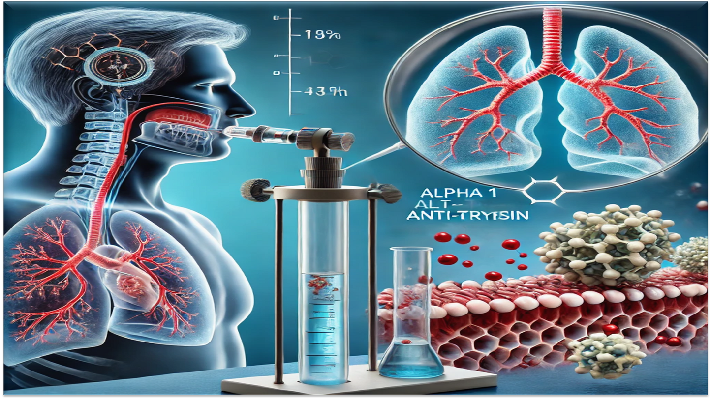
The role of the respiratory tract in paraneoplastic pemphigus has been thoroughly explored, although evidence on pulmonary function in PV is lacking. The current study aims to assess the respiratory function of patients with severe, long-standing PV prior to treatment, as well as to quantify alpha 1-antitrypsin (1-AT) serum levels, in order to identify potential risk factors for lung damage in pemphigus vulgaris patients.
Patients and methods
The institutional Ethical Committee approved this case control study, and patients and controls provided written informed consent. Ten biopsy-proven moderate to severe pemphigus vulgaris patients and ten age-matched controls were enrolled, and data was gathered over a 6-month period on predesigned Performa.
The study included ten patients, four females and six males, who had a positive tzanck test for acantholytic cells with flaccid vesicles and erosions and were hospitalized to the Dermatology Department at Lahore General Hospital. There were no concurrent autoimmune diseases, cardiac or renal abnormalities, or a history of asthma or allergies in the research group.
The study excluded pregnant and breastfeeding women, known smokers, individuals with hereditary alpha 1 deficiency, and patients with a history of tuberculosis or connective tissue illnesses. All of the patients had severe oral lesions. All of the participants in the research had pemphigus vulgaris for more than five years. Pulmonary function tests and alpha 1 antitrypsin level were done on both the study and control groups. The assessment of total lung capacity, vital capacity, and FEV1 was done by pulmonary function testing.
The sick group and the healthy controls' values were compared using the One-Sample Kolmogorov-Smirnov test. P-value (p) was evaluated using SPSS.
Results
Anthropometric traits, lifestyle choices, and physical activity levels were essentially comparable between groups (Figure 1). Out of 10 study participants in case Group showed 6 women and 4 men with ages ranging from 28 to 50, there was a decrease in FEV1 in 4 of the participants, while there was a decrease in alpha 1 antitrypsin in 2 of the participants. While none of the patients in the control group displayed a decrease in alpha 1 antitrypsin level, two patients' levels were reduced when alpha 1 antitrypsin was quantified.
Figure 1 Anthropometric traits and lifestyle comparison between groups, place in results section.
Anthropometric traits, lifestyle choices, and physical activity levels were essentially comparable between groups. Table provides a summary of our findings. Alpha 1 Antitrypsin level was lowered in two patients out of ten pemphigus patients, while significant change was seen in Fev1 in four out of ten pemphigus patients to control to be wither. One patient's HRCT revealed a granuloma in the apico-posterior section of the left upper lobe. One patient had a calcific lung nodule in the right upper lobe, whereas the other two had chronic inflammatory alterations. Another study participant had bilateral epical pleural thickening. These changes support our idea that pulmonary diseases can emerge in pemphigus vulgaris patients. The control group had 3 females and 7 males with an age range of thirty to fifty years all of them showed normal alpha 1 antitrypsin level, normal lung functions and their HRCT chest also turned out to be normal.
The two group were assessed i.e. cases of pemphigus vulgaries and age matched healthy control, for mean value of FEV1 and Alpha1 Antitrypsin. The mean values of FEV1 were calculated in both groups in liters per minute and independent samples t-test applied for the significance among cases and controls, turned out to be 0.0017 i.e. <0.05. The demographic and clinical characteristics of cases and controls are summarized in Table 1. For Alpha1 Anti trypsin the mean values among cases and controls compared with each other applying same independent samples t-test found to 0.001 i.e. <0.05.
Table 1 Characteristics summary of cases and controls, place after patient demographics.
|
Cases Controls Total |
Age
≤ 50 years 3 (75%) 1 (25%) 4
≥50 years 7 (43.8%) 9 (56.3%) 16
Sex
Females 6 (66.7%) 3 (33.3%) 9
Males 4 (36.4%) 7 (63.6%) 11
Other diseases
Hypertension 2 (40%) 3 (60%) 5
Duodenal ulcer 1 (100%) Nil 1
History of pleurisy 2 (100%) Nil 2
Pulmonary disease 1 (100%) Nil 1
Renal lithiasis 1 (100%) Nil 1
Seasonal rhinitis 1 (100%) Nil 1
None 2 (22.2%) 7 (77.8%)
Pemphigus disease activity is noted in study group using the pemphigus disease area index which includes the following proformas. This scoring system includes a score ranging from 0-263, which comprises of 250 points representing disease activity and 120 points for skin activity 10 points for scalp activity and 120 points for mucosal activity.
Extent of disease according to PDAI
Moderate 0-15 points
Significant 15-45 points
Extensive >45 points
Discussion
In our study 40% Pemphigus vulgaris patients had lung changes including infections while in a study of Belgnaoui, et al, 68% had lung changes with infections. Our results are comparable and small differences between two studies may due to differences in severity of disease and duration of hospitalization. Pulmonary findings, including granulomas and pleural thickening, are illustrated in Figure 2. The study by Ljubojevic S et al. on 159 PV Patients, during 19 years revealed several complications associated with high doses of corticosteroids and immunosuppressive
Table 2 PDAI scoring and disease activity, place in results section.
|
PDAI |
Patient number in study group |
|
Moderate |
4 |
|
Severe |
4 |
|
Extensive |
2 |
Figure 2 Pulmonary findings in patients, place in discussion section.
Table 3 Results table of independent samples t-test.
|
Groups |
N |
Mean |
SD |
P-value |
|
Fev1 |
|
|
|
|
|
Cases |
10 |
1.54 |
±0.53 |
0.0017 |
|
Controls |
10 |
2.50 |
±0.20 |
|
|
Alpha 1 anti-trypsin levels |
||||
|
Cases |
10 |
95.00 |
±17.09 |
0.001 |
|
Controls |
10 |
190.0 |
±27.23 |
|
therapy, including Skin infections in 16.35%, sepsis in 5.66% and 14 patients (8.81%) died during the period of hospitalization. The absence of sepsis and death in our study may be due to small number of patients with severe PV and shorter period of our study.6
The extent of disease severity, as assessed using the PDAI scoring system, is detailed in Table 2. Pemphigus vulgaris affects stratified squamous epithelium. So far, there are no clinical trials evaluating possible factors that could enhance susceptibility of PV patients to pulmonary infections. Upper and lower respiratory epithelial cells express all the planking antigens. Specifically paraneoplastic pemphigus is classically related to bronchiolitis obliterans. However, there is no evidence that antibodies against desmogliens play any part in the induction of respiratory lesions. These results coherent with the study conducted by Nguyen VT in 2001.7
The results of the independent samples t-test for FEV1 and alpha 1 antitrypsin level are presented in Table 3. In our study, patients in study group with age above 50 were 7 and in control group age above 50 were 9. 14 were female patients in total, both Pemphigus and control group i.e.70 percent and 6 Male in total i.e. 30 percent. Fev1 in Pemphigus group was found to be reduced, 4 patients, 3 females and 1 Male with mean 1.54 liters SD ±0.53 and in control group all cases had FEV1 within normal range with mean value of 2.5 liters ±0.20 after applying independent t-test on both groups P value calculated P <0.0017. Alpha 1 Antitrypsin levels were found to be reduced in 2 patients in study group with mean of 95.0±17.09 and in control group was 190.0±27.23. P<0.001 showing significant difference It is believed that insufficient production, or prolonged consumption of Alpha 1 might predispose these patients for lung infection and for chronic obstructive lung disease. These results are coherent with the study conducted by Holme J, Stockley Rain 2007.8
According to studies, the rise of anti-proteases like 1-AT causes the proteolytic activity of blister fluid in PV to gradually diminish. According to recent study, matrix metalloproteinase-9 (MMP-9) in skin tissue is inhibited by 1-AT and 1 antichymotrypsin. MMP-9 is reported to be over expressed in experimental PV models. Blister formation in PV may be influenced by an imbalance in the concentrations of MMP-9 and 1-AT. These results are coherent with our results.9
Conclusion
This study concludes that lung changes do occur in cases of pemphigus vulgaris with longer duration along with reduction in FEV1 and alpha 1 antitrypsin level in a significant manner.
Declaration of patient consent the authors certify that they have obtained all appropriate patient consent.
Financial support and sponsorship None.
Conflict of interest Authors declared no conflict of interest.
Authors’ contribution
TK: Substantial contribution to conception and study design, acquisition of data, manuscript writing, has given final approval of the version of the manuscript to be published.
SM: Substantial contribution to analysis and interpretation of data, critical review of the manuscript, has given final approval of the version of the manuscript to be published.
References
1. Kridin K, Sagi SZ, Bergman R. Mortality and cause of death in patients with pemphigus. Acta dermato-venereologica. 2017;97(5):607-11.
2. Di Zenzo G, Amber KT, Sayar BS, Müller EJ, Borradori L, editors. Immune response in pemphigus and beyond: progresses and emerging concepts. Seminars in immunopathology; 2016: Springer.
3. Müller R, Svoboda V, Wenzel E, Müller HH, Hertl M. IgG against extracellular subdomains of desmoglein 3 relates to clinical phenotype of pemphigus vulgaris. Experimental dermatology. 2008;17(1):35-43.
4. Olszewska M, Kolacinska-Strasz Z, Sulej J, Labecka H, Cwikla J, Natorska U, et al. Efficacy and safety of cyclophosphamide, azathioprine, and cyclosporine (ciclosporin) as adjuvant drugs in pemphigus vulgaris. American journal of clinical dermatology. 2007; 8:85-92.
5. Strowd LC, Taylor SL, Jorizzo JL, Namazi MR. Therapeutic ladder for pemphigus vulgaris: emphasis on achieving complete remission. Journal of the American Academy of Dermatology. 2011;64(3):490-4.
6. Ljubojevic S, Lipozenčić J. Autoimmune bullous diseases associations. Clinics in dermatology. 2012;30(1):17-33.
7. Nguyen VT, Ndoye A, Bassler KD, Shultz LD, Shields MC, Ruben BS, et al. Classification, clinical manifestations, and immunopathological mechanisms of the epithelial variant of paraneoplastic autoimmune multiorgan syndrome: a reappraisal of paraneoplastic pemphigus. Archives of Dermatology. 2001;137(2):193-206.
8. Holme J, Stockley RA. Radiologic and clinical features of COPD patients with discordant pulmonary physiology: lessons from α1-antitrypsin deficiency. Chest. 2007;132(3):909-15.
9. Cirillo N, Dell’Ermo A, Gombos F, Lanza A. The specific proteolysis hypothesis of pemphigus: Does the song remain the same? Medical hypotheses. 2008;70(2):333-7.
Share